Folate Receptor Autoantibodies, Multiple Sclerosis, Endometriosis and Myelofibrosis: Are the Causes of Epigenetically Derived Diseases Chronically Neglected?
Different pathologies, different tissues, same mechanism. Follow that mechanism, and a pattern emerges...
BIOLOGYEPIGENETICSSYSTEMS
Alexandra Chambers
5/13/20267 min read
Multiple sclerosis (MS) is usually described as an immune-mediated disease of the central nervous system in which myelin is damaged, producing white-matter lesions and progressive neurological dysfunction. Cerebral folate deficiency/ FRAA (folate receptor autoantibodies) is usually described separately: a disorder of impaired folate transport into the brain, often associated with developmental delay, regression, seizures, behavioural disturbance and abnormal white-matter findings in autistic children.
These conditions sit in different diagnostic categories, and are usually managed by different clinical pathways - but should they be?
A study by Mashayekhi et al. (2024) reported significantly elevated serum folate receptor alpha autoantibodies in patients with relapsing-remitting multiple sclerosis compared with controls. The authors found mean FRAA levels of 67.20 ± 19.79 ng/ml in RRMS patients compared with 37.32 ± 13.26 ng/ml in controls, and concluded that high folate receptor alpha autoantibody concentration was associated with RRMS (Relapsing-Remitting MS).
Folate receptor alpha autoantibodies are already implicated in cerebral folate deficiency, a condition in which folate transport into the cerebrospinal fluid may be impaired even when blood folate is not obviously deficient. FOLR1-related cerebral folate transport deficiency impairs folate transport into CSF and can produce neural folate deficiency without systemic folate deficiency.
This raises an important mechanistic question: if folate receptor autoimmunity is associated with cerebral folate deficiency, and is also elevated in relapsing-remitting MS, should these conditions be identified as separate diseases, or as different expressions of a shared immune-folate-white-matter vulnerability?
Both conditions involve the same neurological territory: white matter, myelin, neural conduction and repair.
In cerebral folate deficiency/FRAA, case reports describe children developing neurological regression, drug-resistant seizures, spasticity and abnormal white-matter signals on brain MRI alongside low CSF (cerebrospinal fluid) 5-MTHF (Methylfolate).
In MS, the target is also white matter and myelin. MS is characterised by immune dysregulation, demyelination, neuroinflammation, axonal injury and progressive neurological disability. White matter lesions have also been noted under MRI.
The diagnostic names differ, but the terrain is strikingly similar: disturbed folate transport can harm white matter; immune-mediated demyelination can harm white matter. The responsible question is therefore why different labelled conditions converge on the same CNS signalling infrastructure.
To say MS is autoimmune or immune-mediated is not wrong, but it is incomplete. It describes the downstream immune pattern, but it does not explain why immune tolerance failed, why myelin became vulnerable, why repair did not resolve the damage, or why one person develops MS while another person exposed to similar stressors does not.
A better model is:
Environmental exposure + divergent genomics + epigenetic immune disruption = tissue-specific disease expression.
In MS, that tissue-specific expression may be demyelination, white-matter lesions, gliosis, axonal injury and failed remyelination. In cerebral folate deficiency/FRAA, the expression may be impaired brain folate transport, abnormal myelination, seizures, regression and white-matter abnormalities. Different labels, but overlapping upstream terrain.
Epigenetics is the bridge. MS research already discusses epigenetic changes in immune cells and oligodendrocyte-lineage cells, meaning that environmental conditions may alter immune behaviour and myelin biology without changing the DNA sequence itself. Something has therefore pushed immune regulation, tolerance, recognition, repair and inflammatory signalling into a pathological state.
Folate receptor alpha is crucial because it sits at a boundary between blood folate status and brain folate availability. A person may have adequate or even high systemic folate whilst having impaired folate delivery into the central nervous system if folate receptor transport is disrupted.
That makes FRAA a particularly important candidate mechanism. It may represent immune interference with folate access to the brain. If unresolved, this could plausibly affect methylation, nucleotide repair, mitochondrial function, oligodendrocyte support, myelin maintenance, immune regulation and neural repair capacity. These are central to whether white matter remains stable, repairs properly, or becomes chronically vulnerable.
The progression hypothesis would be:
Folate receptor autoantibodies → impaired cerebral folate transport → reduced CNS folate availability → methylation and repair disturbance → oligodendrocyte/myelin vulnerability → abnormal white-matter signal or impaired myelination → inflammation, failed repair, gliosis or demyelinating progression in susceptible individuals.
This is not proof that FRAA causes MS, but the 2024 RRMS/FRAA study means the hypothesis is no longer merely speculative. FRAA has now appeared inside the MS conversation and should be investigated properly.
A child with folate transport disturbance, seizures, developmental regression, autistic traits or white-matter abnormalities may be placed in a neurodevelopmental or metabolic category. An adult with demyelinating lesions, sensory symptoms, fatigue, vision disturbance or motor dysfunction may be placed in an MS category.
What if they are different temporal windows of a shared vulnerability though?
Early-life expression may look like:
Developmental delay, autism-associated features, ADHD-like dysregulation, epilepsy, regression, behavioural disturbance, hypotonia, movement disorder or abnormal myelination.
Later-life expression may look like:
Relapsing-remitting MS, demyelination, white-matter lesions, fatigue, sensory disturbance, motor dysfunction, cognitive changes, gliosis, failed remyelination or progressive neurodegeneration.
The label changes, but the tissue vulnerability may not.
This is where synthetic folic acid becomes difficult to ignore. It cannot be blamed as the only possible cause of MS, cerebral folate deficiency or neurodevelopmental harm, that would likely be too reductionist. However, environmental pollutants, heavy metals, lead, mercury, solvents, pesticides, infections, trauma physiology, pharmaceutical exposures, endocrine disruption, nutritional depletion and chronic inflammatory load all belong in the wider integration of the exposome.
The synthetic nutrient folic acid (sometimes listed as B9 on food packaging) does keep reappearing because folate biology is central to the exact systems being discussed: methylation, immune regulation, neurodevelopment, myelin maintenance, mitochondrial function, DNA repair and CNS folate transport.
Synthetic folic acid is not the same as naturally occurring food folates or reduced folate forms such as folinic acid. Folic acid can bind folate receptor alpha very strongly, creating a problematic substrate, especially when exposed frequently to processed foods - and this interferes with natural folate transport.
Folic acid may therefore be one modern convergence node in a broader immune-folate-myelin harm pathway, especially where folate receptor autoimmunity, divergent genomics, impaired methylation, neurodevelopmental vulnerability or white-matter abnormalities are already present.
Neurodivergence also belongs in this conversation because (neuro)divergent populations carry divergent genomics, immune, metabolic, sensory, connective-tissue, folate-handling, glutamate/GABA and mitochondrial profiles that alter how environmental pressures are absorbed and expressed.
(Also, many (neuro)divergents are historically - and currently - undiagnosed and unrecognised).
In one person, the terrain may express as emotional dysregulation, sensory overload and epilepsy. In another, it may express as connective tissue instability, autoimmune disease, gut inflammation or migraine. In another, the vulnerable endpoint may be myelin and white matter.
This is why the same mechanistic themes keep returning: folate, glutamate, epilepsy, white matter, immune misdirection, methylation, connective tissue, oxidative stress, mitochondrial load and environmental exposure. They are not random fragments; they are all part of the same wider question:
What happens when genomically divergent populations are repeatedly exposed to environmental pressures that disrupt immune regulation, methylation, repair and neurological signalling? MS may be one answer.
Endometriosis and myelofibrosis
Endometriosis and myelofibrosis are not the same disease. However, they may also be different tissue-specific expressions of the same underlying harm, or at least the same class of harm.
Both involve fibrotic remodelling: a state in which tissue does not return cleanly to normal after injury or disruption, but instead shifts into abnormal persistence, structural change, and progressive dysfunction. Once that happens, the bod is remodelling along a pathological path.
That process will not look the same in every tissue. Tissue shapes the outcome; cell type, blood supply, immune behaviour, hormonal environment, local signalling, mechanical stress, and structural role all affect how harm is expressed. The same broad injurious driver may therefore produce very different visible disease depending on where it lands.
In one tissue, the result may be ectopic persistence, inflammation, adhesions, and scarring. In another, it may be marrow fibrosis, disrupted blood formation, and gradual loss of normal tissue function. The presentation differs because the tissue differs. That does not rule out a shared underlying cause. In fact, this may be exactly what we would expect if the same harmful process were being filtered through different biological environments.
A condition is usually named according to where it is found, how it presents, and what it looks like by the time medicine recognises it. That does not necessarily tell us whether the initiating harm was unique to that condition. It only tells us how the body expressed the damage in that location, and how medicine happened to encounter it.
The timing of recognition matters here as much as the tissue itself. Conditions do not all force themselves into a diagnostic view in the same way. Some are recognised earlier because their symptoms are harder to ignore, more disruptive, or more obviously abnormal. Others remain less visible for longer, because they do not announce themselves as clearly in the early stages.
That difference changes how medicine perceives them. Endometriosis is more likely to come to attention through pain, bleeding, infertility, bowel disturbance, or other disruptive symptoms. Myelofibrosis may remain less obvious for much longer and only become identifiable once secondary changes are more established. That means the two conditions may not only be occurring in different tissues. They may also be entering medicine at different stages of progression.
That alone can make them appear far more different than they really are.
If one process is usually recognised through an earlier or more symptom-heavy phase, while another is more often picked up later through downstream structural or functional consequences, medicine is not comparing like with like. It is comparing different points along different timelines of visibility. Once that happens, the label begins to harden around the moment of recognition, rather than around the full biological story.
Therefore, the gap between endometriosis and myelofibrosis may not be as wide as it first appears.
If connective tissue architecture, extracellular matrix behaviour, and repair capacity differ between individuals, then it makes sense to ask whether some people are more vulnerable to this kind of pathological remodelling across multiple tissues.
The mechanisms are often visible in the literature, distributed across pathology, immunology, metabolism, endocrinology, genetics, and epigenetics. The problem is that they are rarely integrated into a causal framework. Each paper may describe part of the mechanism, each specialty may observe part of the injury, yet the system as a whole avoids the conclusion that would follow from joining them together.
This is a significant form of epistemic negligence: a failure to know what the evidence already makes knowable. Intent does not need to be proven for harm to be recognised. If the causal conditions of disease are repeatedly described, yet prevention is not pursued, the result is not neutral. The harm continues because the mechanism is observed, named in fragments, and then left unconnected.
Study DOI: 10.1016/j.clineuro.2024.108161
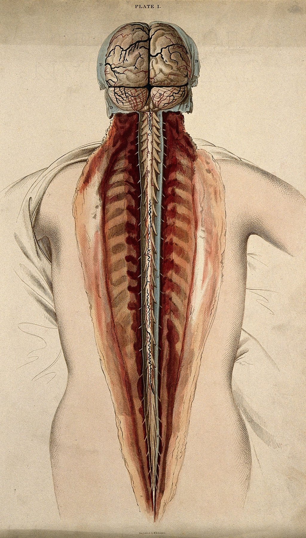
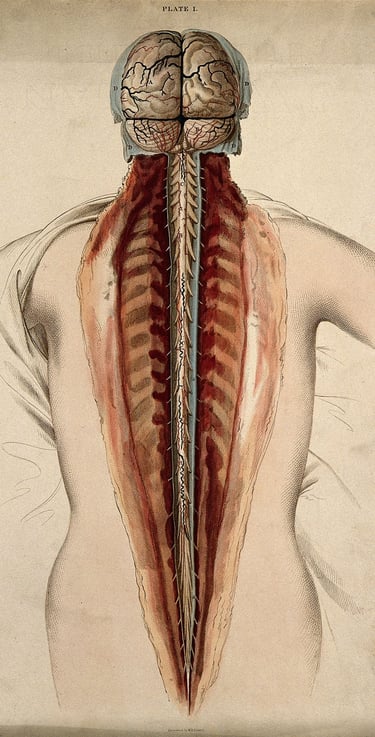
Artist: W.H. Lizars, ca. 1827
Contact
Reach out with questions or collaboration ideas.
AChambers@divergentgenomics.org
© Alexandra Chambers 2026. All rights reserved.